File:Morbus Fabry MRT Osteoporosis 01.jpg
Jump to navigation
Jump to search

Size of this preview: 800 × 490 pixels. Other resolutions: 320 × 196 pixels | 640 × 392 pixels | 1,200 × 735 pixels.
Original file (1,200 × 735 pixels, file size: 147 KB, MIME type: image/jpeg)
Captions
Captions
Add a one-line explanation of what this file represents
| Description |
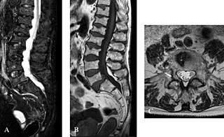
English: Bone magnetic resonance imaging in a Fabry patient with severe osteoporosis: A (STIR, sagittal view) and B (T1, sagital median): several vertebral body fractures are seen, without signal anomaly in T1 or T2 in favor of ancient fractures. A mild spondylolisthesis of L5 on S1 can be observed. C (T2, axial view): fracture of the right pedicula of L5 (arrow) in a 72-year-old patient with severe osteoporosis. Courtesy: Dr Robert CARLIER, CHU Raymond Poincaré, Garches, France.
Deutsch: Magnetresonanztomographie eines Morbus-Fabry-Patienten mit schwerer Osteoporose. (A) Sagittale Betrachtung. Gemessen mit einer STIR-Sequenz (Short-Tau Inversion Recovery), (B) T1-gewichtet, sagittal mediane Betrachtung. Eine Reihe von Frakturen der Wirbelkörper sind sichtbar. Eine leichte Spondylolisthesis von L5 auf S1 kann ebenfalls diagnostiziert werden. (C) Eine T2-gewichtete axiale Aufnahme zeigt eine Fraktur (Pfeil) des rechten Füßchens des Wirbelbogens (Pediculus arcus vertebrae) von L5, bei einem 72-Jährigen Patienten mit schwerer Osteoporose. |
| Date | article published: 22 November 2010 [1] |
| Source | D. P. Germain: Fabry disease. In: Orphanet journal of rare diseases Vol. 5, 2010, 30, PMID 21092187. PMC 3009617. (Review) |
| Author | Dr Robert CARLIER, CHU Raymond Poincaré, Garches, France. |
This file is licensed under the Creative Commons Attribution 2.0 Generic license.
- You are free:
- to share – to copy, distribute and transmit the work
- to remix – to adapt the work
- Under the following conditions:
- attribution – You must give appropriate credit, provide a link to the license, and indicate if changes were made. You may do so in any reasonable manner, but not in any way that suggests the licensor endorses you or your use.
File history
Click on a date/time to view the file as it appeared at that time.
| Date/Time | Thumbnail | Dimensions | User | Comment | |
|---|---|---|---|---|---|
| current | 12:05, 10 September 2011 | | 1,200 × 735 (147 KB) | Kuebi (talk | contribs) | {{Information |Description={{en|Bone magnetic resonance imaging in a Fabry patient with severe osteoporosis: A (STIR, sagittal view) and B (T1, sagital median): several vertebral body fractures are seen, without signal anomaly in T1 or T2 in favor of anci |
You cannot overwrite this file.
File usage on Commons
The following 3 pages use this file:
File usage on other wikis
The following other wikis use this file:
- Usage on de.wikipedia.org
- Usage on en.wikipedia.org
- Usage on en.wikibooks.org
- Usage on outreach.wikimedia.org
- Usage on sr.wikipedia.org
- Usage on uz.wikipedia.org