Category:Media from BioMed Central needing categories
Jump to navigation
Jump to search
This category contains files originating from BioMed Central that have not yet been categorized by journal. This categorization should be performed as in this example. For an overview of existing categories, see Category:Media from BioMed Central.
Media in category "Media from BioMed Central needing categories"
The following 133 files are in this category, out of 133 total.
-
 Double fertilization in arabidopsis 2.jpg 1,499 × 652; 234 KB
-
 Double fertilization in arabidopsis.jpg 1,200 × 977; 376 KB
-
 Double immunofluorescence staining for BrdU, NeuN and GFAP.jpg 1,200 × 1,087; 363 KB
-

-
 DuPanSyndrome2.jpg 400 × 267; 10 KB
-
 E amelogenesis imperfecta.jpg 360 × 260; 19 KB
-
 Eccrine hydrocystoma 01.jpg 1,200 × 1,793; 481 KB
-
 Edema Hands 01.jpg 1,013 × 646; 287 KB
-
 Electron microscopy reveals mitochondrial DNA in discrete foci.jpg 956 × 1,236; 248 KB
-
 Eosinophilic myocarditis HE stain.jpg 621 × 618; 46 KB
-
-
 Events in alpha synuclein toxicity.jpg 1,200 × 1,250; 281 KB
-
 F amelogenesis imperfecta.jpg 360 × 260; 22 KB
-

-
 Friedreich.jpg 410 × 612; 36 KB
-
 Fundal photograph showing severe papilloedema in the right eye.jpg 640 × 480; 58 KB
-
 G-CSF receptor is expressed in the embryonic nervous system radial glia.jpg 1,200 × 1,055; 592 KB
-
 Gabaergic inhibition by chandelier cells.jpg 1,200 × 1,174; 155 KB
-
 Gad1 transcripts in the developing vibrissae.jpg 1,200 × 2,018; 131 KB
-
 Ganglionic eminence mice E12.5 doi 10.1186 1747-5333-1-16-1.jpg 1,200 × 900; 177 KB
-
 Gas gangrene pathology slide.jpg 1,038 × 771; 100 KB
-
 Gas gangrene.jpg 1,200 × 960; 245 KB
-
 Gelatinous drop-like corneal dystrophy 1.JPEG 499 × 499; 98 KB
-
 Gelatinous drop-like corneal dystrophy 2.JPEG 500 × 500; 110 KB
-
 Gelatinous drop-like corneal dystrophy 3.JPEG 500 × 500; 76 KB
-
 Gelatinous drop-like corneal dystrophy 4.JPEG 499 × 499; 78 KB
-
 Gelatinous drop-like corneal dystrophy 5.JPEG 500 × 328; 99 KB
-
 Genomic organization of CPLX2 and locations of SNPs.png 617 × 272; 44 KB
-
 Genomic organization of SYN2 and locations of SNPs.png 1,140 × 284; 71 KB
-
 Girl with Noonan syndrome.jpg 866 × 742; 43 KB
-
 Glial distribution of Cu Zn SOD immunoreactivity in intact immature rat brain.jpg 1,200 × 1,222; 577 KB
-

-
 Gliomatosis cerebri2.jpg 1,200 × 1,499; 284 KB
-

-
-
 Heart kabuki syndrome.jpg 1,200 × 804; 177 KB
-
 Hemangioma 01.jpg 1,200 × 808; 249 KB
-
 Hemipelvectomy gas gangrene.jpg 1,200 × 800; 288 KB
-
 Hemochromatosis liver iron prussian blue.jpg 1,200 × 960; 395 KB
-
 Hemoscrotum.JPEG 2,272 × 1,704; 930 KB
-
 Hepatic stellate cell (ito cell) 1476-5926-6-7-3-l.jpg 423 × 687; 178 KB
-
 Hyperostosis.jpg 1,200 × 900; 85 KB
-
 Integrated Model to Assess the Global Environment (IMAGE 2) model schematic.jpg 1,200 × 1,900; 206 KB
-

-
 Kaposis sarcoma 01.jpg 1,200 × 809; 266 KB
-
 Kawasaki symptoms A.jpg 464 × 384; 19 KB
-
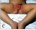 Kawasaki symptoms C.jpg 464 × 384; 19 KB
-
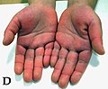 Kawasaki symptoms D.jpg 448 × 368; 18 KB
-
 Kawasaki symptoms E.jpg 464 × 368; 18 KB
-
 Kawasaki symptoms F.jpg 464 × 368; 22 KB
-
 Kawasaki symptoms G.jpg 464 × 368; 21 KB
-
 Kawasaki symptoms H.jpg 464 × 368; 21 KB
-
 Kawasaki.PNG 1,900 × 767; 1.77 MB
-
-
 Langerhans cell histiocytosis of the bone.jpg 1,200 × 900; 253 KB
-
 Laron syndrome 01.jpg 1,109 × 812; 245 KB
-
 Lattice corneal dystrophy type 1.JPEG 500 × 500; 71 KB
-
 Lattice corneal dystrophy type II cornea cut.jpg 701 × 549; 149 KB
-
 Lattice corneal dystrophy type II diagram.gif 1,271 × 845; 340 KB
-
 Leg Edema 01.jpg 591 × 403; 105 KB
-
 Leg Edema 02.jpg 599 × 403; 99 KB
-
 Leiomyosarcoma of the Adrenal vein 1.jpg 1,200 × 1,004; 480 KB
-
 Life cycle and protein associations of connexins.jpg 1,200 × 1,442; 230 KB
-

-
 Liver, high power, demonstrating Cowdry bodies within hepatocytes.jpg 1,200 × 900; 243 KB
-

-
 Macular corneal dystrophy - GAG killed keratocyte.JPEG 500 × 500; 109 KB
-
 Macular corneal dystrophy hale colloidal iron stain.JPEG 500 × 500; 152 KB
-
 Macular corneal dystrophy lamp exam.JPEG 500 × 500; 66 KB
-
 Major cellular sources of Reactive Oxygen Species in living cells.jpg 1,200 × 1,600; 227 KB
-
 Major metabolic fluxes in neuron-astrocyte coupling for resting conditions.png 1,024 × 710; 330 KB
-

-

-

-
 Metaphase spread of the Siberian Roe deer (Capreolus pygargus).jpg 391 × 452; 26 KB
-

-
 Metaphase spread of the Viscacha rat (Tympanoctomys barrerae).jpg 404 × 385; 22 KB
-
 Morbus Fabry aortic root dilatation 01.jpg 1,200 × 1,182; 118 KB
-
 Morbus Fabry aortic root dilatation 02.jpg 1,200 × 740; 89 KB
-
 Morbus Fabry Doppler 01.jpg 1,200 × 836; 156 KB
-
 Morbus Fabry DXA 01.jpg 1,200 × 763; 137 KB
-
 Morbus Fabry EKG 02.jpg 1,200 × 549; 130 KB
-
 Morbus Fabry ganglion cells.jpg 1,200 × 687; 321 KB
-
 Morbus Fabry Genotyping 01.jpg 1,200 × 896; 247 KB
-
 Morbus Fabry Hypoacousia 01.jpg 1,200 × 1,310; 237 KB
-
 Morbus Fabry kidney biopsy 01.jpg 1,181 × 794; 342 KB
-
 Morbus Fabry kidney biopsy TEM 01.jpg 1,181 × 821; 365 KB
-
 Morbus Fabry kidney biopsy TEM 02.jpg 1,181 × 829; 448 KB
-
 Morbus Fabry kidney biopsy TEM 03.jpg 1,181 × 822; 450 KB
-
 Morbus Fabry LVH echo 01.jpg 1,200 × 829; 87 KB
-
 Morbus Fabry MRA 01.jpg 1,200 × 999; 172 KB
-
 Morbus Fabry MRI 01.jpg 1,200 × 428; 65 KB
-
 Morbus Fabry MRT Osteoporosis 01.jpg 1,200 × 735; 147 KB
-
 Morbus Fabry pulvinar sign 01.jpg 1,200 × 1,339; 302 KB
-
 Morbus Fabry Skin Rash 01.jpg 1,200 × 914; 122 KB
-
 Morbus Fabry Stroke MRT 01.jpg 1,200 × 678; 125 KB
-
 Morbus Fabry Tissue Doppler 01.jpg 1,200 × 833; 110 KB
-
 Morbus Osler 01.jpg 1,200 × 794; 448 KB
-
 Mup locus showing DNA repeats.jpg 582 × 686; 153 KB
-

-
 Myasthenia gravis ptosis reversal.jpg 1,160 × 260; 133 KB
-
 Naxos Disease Hair 01.jpg 585 × 516; 203 KB
-
 Naxos Disease Hand Feet 01.jpg 564 × 636; 404 KB
-
 Naxos-Syndrom EKG 01.jpg 1,200 × 338; 147 KB
-
 Naxos-Syndrom HEstain 01.jpg 1,200 × 876; 233 KB
-
 Necrotizing fasciitis left leg debridement.JPEG 2,132 × 1,470; 142 KB
-
 Necrotizing fasciitis left leg.JPEG 2,288 × 1,313; 135 KB
-
 Neurogenesis and rTMS.jpg 1,200 × 551; 115 KB
-
 Nevus araneus 01.jpg 1,200 × 782; 327 KB
-
 NOGO-A expression during cell migration.jpg 1,200 × 605; 395 KB
-
 Noonan syndrome.PNG 753 × 645; 367 KB
-
 Nuclear envelope breakdown and reassembly in mitosis.jpg 1,200 × 790; 142 KB
-
 Nuclear integrity and genome stability in normal and HGPS cells.jpg 600 × 723; 64 KB
-
 Oesophagusatresie - Einteilung nach Vogt.png 581 × 250; 172 KB
-
 Penile Edema 01.jpg 597 × 413; 171 KB
-
 Penile Edema 02.jpg 602 × 412; 95 KB
-
 Rosacea 01.jpg 1,200 × 793; 337 KB
-
 Schematic drawing of cellular regulation of extracellular glutamate concentrations.jpg 1,200 × 2,099; 415 KB
-
 Schematic Examples of CNS Structural Changes in chronic pain.jpg 1,200 × 310; 90 KB
-

-
 Schematic of cortical areas involved with pain processing and fMRI cropped.jpg 1,172 × 792; 211 KB
-
 Schematic of cortical areas involved with pain processing and fMRI.jpg 1,200 × 1,383; 378 KB
-

-
 Schematic representation of the human collagen XVIII variants.jpg 1,200 × 312; 44 KB
-

-
 Solitary pulmonary nodule CT arrow.jpg 1,188 × 1,200; 118 KB
-
 Splenic infarction.png 940 × 668; 196 KB
-
 Spondylitis Tropheryma whipplei.jpg 196 × 505; 60 KB
-
 Spore germination in Saccharomyces cerevisiae.png 534 × 637; 75 KB
-
 Streptococcus iniae fibrinogen binding.png 2,126 × 703; 1.55 MB
-
 Streptococcus iniae.png 350 × 271; 131 KB
-
 Structure and function of the nuclear lamina.jpg 1,200 × 593; 133 KB
-
 Structure of NR4A1 bound to DNA.png 482 × 332; 178 KB


_factors_regulating_their_cytoskeletal_dynamics.jpg)


_1476-5926-6-7-3-l.jpg)

_model_schematic.jpg)


.jpg)


.jpg)
.jpg)
.jpg)


.jpg)