File:A-workflow-to-process-3D+time-microscopy-images-of-developing-organisms-and-reconstruct-their-cell-ncomms9674-s12.ogv
Jump to navigation
Jump to search
Size of this JPG preview of this OGG file: 800 × 478 pixels. Other resolutions: 320 × 191 pixels | 640 × 383 pixels | 836 × 500 pixels.
Original file (Ogg Theora video file, length 36 s, 836 × 500 pixels, 3.31 Mbps, file size: 14.04 MB)
Captions
Captions
Add a one-line explanation of what this file represents
Summary[edit]
| Description |
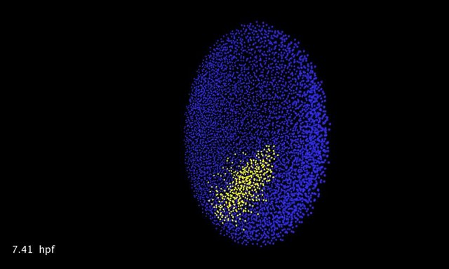
English: Supplementary Movie 11 Three cell populations identified according to their position and morphogenetic movements: epiblast cells (in blue), hypoblast cells (in yellow) entering the imaged volume by 5.70 hpf, enveloping layer (EVL) cells (in cyan) highlighted between 4.24 hpf and 5.66 hpf by a 3D Delaunay triangulation joining the nucleus of neighbor cells (Mov-IT visualization tool). |
||
| Date | |||
| Source | Video file from Faure E, Savy T, Rizzi B, Melani C, Stašová O, Fabrèges D, Špir R, Hammons M, Čúnderlík R, Recher G, Lombardot B, Duloquin L, Colin I, Kollár J, Desnoulez S, Affaticati P, Maury B, Boyreau A, Nief J, Calvat P, Vernier P, Frain M, Lutfalla G, Kergosien Y, Suret P, Remešíková M, Doursat R, Sarti A, Mikula K, Peyriéras N, Bourgine P (2016). "A workflow to process 3D+time microscopy images of developing organisms and reconstruct their cell lineage". Nature Communications. DOI:10.1038/ncomms9674. PMID 26912388. PMC: 4773431. | ||
| Author | Faure E, Savy T, Rizzi B, Melani C, Stašová O, Fabrèges D, Špir R, Hammons M, Čúnderlík R, Recher G, Lombardot B, Duloquin L, Colin I, Kollár J, Desnoulez S, Affaticati P, Maury B, Boyreau A, Nief J, Calvat P, Vernier P, Frain M, Lutfalla G, Kergosien Y, Suret P, Remešíková M, Doursat R, Sarti A, Mikula K, Peyriéras N, Bourgine P | ||
| Permission (Reusing this file) |
This file is licensed under the Creative Commons Attribution 4.0 International license.
|
||
| Provenance |
|
File history
Click on a date/time to view the file as it appeared at that time.
| Date/Time | Thumbnail | Dimensions | User | Comment | |
|---|---|---|---|---|---|
| current | 01:16, 29 October 2016 | 36 s, 836 × 500 (14.04 MB) | Open Access Media Importer Bot (talk | contribs) | Automatically uploaded media file from Open Access source. Please report problems or suggestions here. |
You cannot overwrite this file.
File usage on Commons
There are no pages that use this file.