Category:Cytological techniques
Jump to navigation
Jump to search
| Upload media | |||||
| Instance of |
| ||||
|---|---|---|---|---|---|
| Subclass of |
| ||||
| Different from | |||||
| |||||
Subcategories
This category has the following 3 subcategories, out of 3 total.
A
C
- Cell fractionation (7 F)
Media in category "Cytological techniques"
The following 60 files are in this category, out of 60 total.
-
A-distributed-cell-division-counter-reveals-growth-dynamics-in-the-gut-microbiota-ncomms10039-s2.ogv 6.2 s, 623 × 623; 5.62 MB
-
-
-
-
-
-
-
-
-
-
-
-
-
-
-
-
-
-
-
-
Automated-measurement-of-cell-motility-and-proliferation-1471-2121-6-19-S1.ogv 38 s, 320 × 240; 2.43 MB
-
Automated-measurement-of-cell-motility-and-proliferation-1471-2121-6-19-S2.ogv 1 min 19 s, 320 × 240; 687 KB
-
Automated-measurement-of-cell-motility-and-proliferation-1471-2121-6-19-S3.ogv 1 min 51 s, 720 × 480; 1.23 MB
-
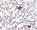
 Basophil in blood.jpg 2,464 × 2,056; 4.11 MB
-
 Burkitt leukemia with cytochemical profile.jpg 2,464 × 2,056; 4.06 MB
-
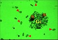
 Contagious cancer.png 2,184 × 1,210; 4.6 MB
-
-
-
-
-
Detection-of-Heteroplasmic-Mitochondrial-DNA-in-Single-Mitochondria-pone.0014359.s003.ogv 1 min 19 s, 960 × 540; 3.39 MB
-
-
Fluorescence-Cell-Imaging-and-Manipulation-Using-Conventional-Halogen-Lamp-Microscopy-pone.0031638.s007.ogv 1 min 51 s, 320 × 240; 860 KB
-
 Glass slide.jpg 3,024 × 4,032; 4.01 MB
-

-
 Kaira alba, broyat de glande agrégée.jpg 5,553 × 3,825; 872 KB
-
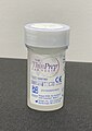 Liquid based cytology specimen pot.jpg 1,691 × 2,397; 1.37 MB
-
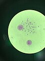
 Metaphase1.jpg 4,032 × 3,024; 1.43 MB
-
 Metaphase3.jpg 3,024 × 4,032; 1.49 MB
-
 Neutrophils with segmented nuclei.jpg 2,464 × 2,056; 3.15 MB
-
 NewStainc.jpg 800 × 224; 48 KB
-
 Red blood cells dispoiesis.jpg 2,464 × 2,056; 3.14 MB
-
-
-
-
Simple-Display-System-of-Mechanical-Properties-of-Cells-and-Their-Dispersion-pone.0034305.s002.ogv 5.7 s, 640 × 480; 231 KB
-
Single-Cell-Analysis-of-Transcriptional-Activation-Dynamics-pone.0010272.s005.ogv 4.4 s, 196 × 187; 45 KB
-
Single-Cell-Analysis-of-Transcriptional-Activation-Dynamics-pone.0010272.s006.ogv 3.6 s, 660 × 155; 129 KB
-
Single-Cell-Analysis-of-Transcriptional-Activation-Dynamics-pone.0010272.s007.ogv 3.6 s, 645 × 215; 472 KB
-
Single-Cell-Analysis-of-Transcriptional-Activation-Dynamics-pone.0010272.s008.ogv 3.6 s, 600 × 200; 167 KB
-
Single-Cell-Analysis-of-Transcriptional-Activation-Dynamics-pone.0010272.s009.ogv 4.0 s, 654 × 212; 209 KB
-
Single-Cell-Analysis-of-Transcriptional-Activation-Dynamics-pone.0010272.s010.ogv 3.6 s, 555 × 165; 258 KB
-
 T.S. of ovary of Hibiscus Double Red Flower.jpg 2,144 × 2,144; 1,014 KB
-
The dance of the ciliated cells - Matteo Gelardi.webm 2 min 47 s, 1,280 × 720; 114.42 MB
-
The-Diverse-and-Dynamic-Nature-of-Leishmania-Parasitophorous-Vacuoles-Studied-by-Multidimensional-pntd.0001518.s003.ogv 1 min 0 s, 364 × 320; 1.65 MB
-
-
The-Diverse-and-Dynamic-Nature-of-Leishmania-Parasitophorous-Vacuoles-Studied-by-Multidimensional-pntd.0001518.s005.ogv 1 min 19 s, 364 × 348; 7.38 MB
-
-
The-Diverse-and-Dynamic-Nature-of-Leishmania-Parasitophorous-Vacuoles-Studied-by-Multidimensional-pntd.0001518.s007.ogv 1 min 3 s, 364 × 296; 1.74 MB
-


.jpg)


