File:Whole-organ thymus perfusion and decellularisation.webp
Jump to navigation
Jump to search

Size of this PNG preview of this WEBP file: 673 × 599 pixels. Other resolutions: 270 × 240 pixels | 539 × 480 pixels | 863 × 768 pixels | 1,150 × 1,024 pixels | 1,998 × 1,779 pixels.
Original file (1,998 × 1,779 pixels, file size: 652 KB, MIME type: image/webp)
Captions
Captions
From the study "Reconstitution of a functional human thymus by postnatal stromal progenitor cells and natural whole-organ scaffolds"
Summary[edit]
| Description |
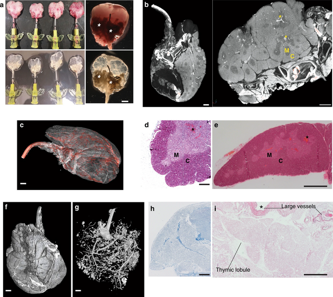
English: "a Gross appearance of cannulated rat thymi before (upper panels) and after (lower panels) decellularisation. A 24G cannula is inserted into the carotid artery and used to perfuse the organ with detergent and enzymatic solutions. Asterisks indicate extra-thymic tissues that allowed connection between thymic tissue and cannula through the large blood vessels (n = 120). Scale bar, 2 mm. b Micro-CT images of cannulated rat thymus showing extra-thymic tissues, large blood vessels and the 24G cannula entering the artery. Iodine contrast shows clear demarcation between cortex (C, bright) and medullary (M) areas; blood vessels (asterisks) are represented by very bright areas between and inside the parenchyma (n = 3). Scale bar, 1.5 mm. c Micro-CT 3D image of whole rat thymus cannulated where iodine contrast shows vasculature tree (segmented in red, n = 2). Scale bar, 1.2 mm. d Masson’s trichrome stain of a fresh rat thymus lobe staining in red keratins, in blue collagen and in pink cytoplasm. C cortex, M medulla (n = 3 thymi). Scale bar, 250 μm. e Haematoxylin & Eosin (H&E) stain of a fresh rat thymus. C cortex, M medulla (n = 3 thymi). Scale bar, 500 μm. f Micro-CT image of cannulated rat thymus showing the whole 2 lobes in 3D. Scale bar, 1.2 mm. g Micro-CT image of a cannulated rat thymus injected with Microfil® and thresholded to demonstrate perfusion of both thymic lobes. Scale bar, 1.2 mm. h Masson’s trichrome stain of a paraffin section of a decellularised rat thymus scaffold demonstrating collagen fibres (blue) and absence of keratins, muscle fibres and cell cytoplasm (n = 3 scaffolds). Scale bar, 250 μm. i H&E of a decellularised thymus scaffold showing intact thymic lobule ECM and preservation of both large and small vasculature wall (n = 4 scaffolds). Scale bar, 500 μm." |
| Date | |
| Source | https://www.nature.com/articles/s41467-020-20082-7 |
| Author | Authors of the study: Sara Campinoti, Asllan Gjinovci, Roberta Ragazzini, Luca Zanieri, Linda Ariza-McNaughton, Marco Catucci, Stefan Boeing, Jong-Eun Park, John C. Hutchinson, Miguel Muñoz-Ruiz, Pierluigi G. Manti, Gianluca Vozza, Carlo E. Villa, Demetra-Ellie Phylactopoulos, Constance Maurer, Giuseppe Testa, Hans J. Stauss, Sarah A. Teichmann, Neil J. Sebire, Adrian C. Hayday, Dominique Bonnet & Paola Bonfanti |
Licensing[edit]
This file is licensed under the Creative Commons Attribution 4.0 International license.
- You are free:
- to share – to copy, distribute and transmit the work
- to remix – to adapt the work
- Under the following conditions:
- attribution – You must give appropriate credit, provide a link to the license, and indicate if changes were made. You may do so in any reasonable manner, but not in any way that suggests the licensor endorses you or your use.
File history
Click on a date/time to view the file as it appeared at that time.
| Date/Time | Thumbnail | Dimensions | User | Comment | |
|---|---|---|---|---|---|
| current | 15:28, 28 January 2021 | | 1,998 × 1,779 (652 KB) | Prototyperspective (talk | contribs) | Uploaded a work by Authors of the study: Sara Campinoti, Asllan Gjinovci, Roberta Ragazzini, Luca Zanieri, Linda Ariza-McNaughton, Marco Catucci, Stefan Boeing, Jong-Eun Park, John C. Hutchinson, Miguel Muñoz-Ruiz, Pierluigi G. Manti, Gianluca Vozza, Carlo E. Villa, Demetra-Ellie Phylactopoulos, Constance Maurer, Giuseppe Testa, Hans J. Stauss, Sarah A. Teichmann, Neil J. Sebire, Adrian C. Hayday, Dominique Bonnet & Paola Bonfanti from https://www.nature.com/articles/s41467-020-20082-7 with U... |
You cannot overwrite this file.
File usage on Commons
There are no pages that use this file.