File:Viruses-11-00733-g003 Virophages.png
Jump to navigation
Jump to search

Size of this preview: 453 × 599 pixels. Other resolutions: 181 × 240 pixels | 363 × 480 pixels | 581 × 768 pixels | 1,129 × 1,493 pixels.
Original file (1,129 × 1,493 pixels, file size: 1.85 MB, MIME type: image/png)
Captions
Captions
Add a one-line explanation of what this file represents
Summary[edit]
| Description |
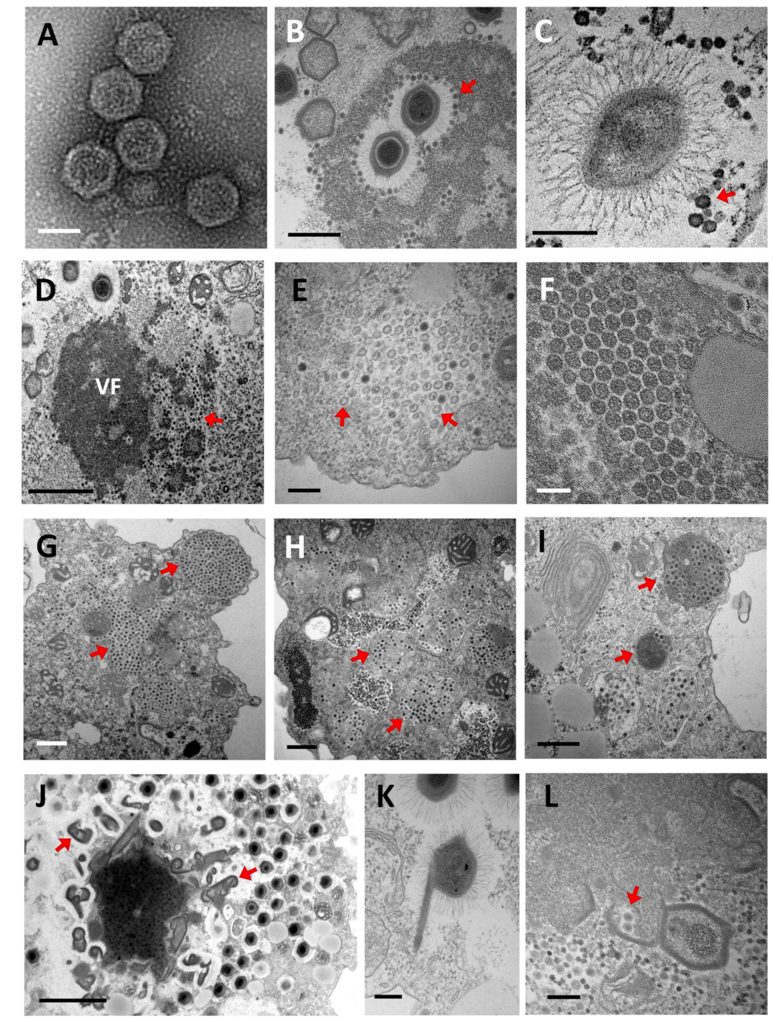
English: Electronic microscopy observation of virophage particles and their replication cycle. (A) Negative staining electronic microscopy observation. (B–L) Transmission electronic microscopy images. (A) Morphology of purified Sputnik virophage particles (scale bar, 500 nm). (B,C) Sputnik and Guarani virions attached to the Mimivirus fibrils, respectively. (Scale bar, 500 nm and 200 nm). (D) Sputnik virophage invading the virus factory of Acanthamoeba polyphaga Mimivirus (APMV) (scale bar, 1 µm). (E) Immature Sputnik virions observed in the cytoplasm of A. castellanii during co-infection with APMV (arrows) (scale bar, 200 nm). (F) Mature Sputnik virions (scale bar, 100 nm). (G–I) Virophages virions are commonly observed clustered inside typical cytoplasmic vesicles at the end of their replication cycles (arrows). (G) Sputnik progeny. (H) Zamilon progeny. (I) Guarani progeny (scale bars, 500 nm). (J,K) The genesis of abnormal Mimivirus particles has been observed during infection with virophages (arrows). (scale bars, 2 µm and 200 nm). (L) Encapsidation of virophage virions within the Mimivirus capsid (arrows) (scale bar, 200 nm). VF: Virus factory. Credit: Said Mougari et al. (2019) |
| Date | |
| Source |
https://www.mdpi.com/viruses/viruses-11-00733/article_deploy/html/images/viruses-11-00733-g003.png at https://www.mdpi.com/1999-4915/11/8/733/htm Virophages of Giant Viruses: An Update at Eleven. Viruses 2019, 11(8), 733; doi:10.3390/v11080733. This article belongs to the Section Viruses of Plants, Fungi and Protozoa. Licensee MDPI, Basel, Switzerland. This article is an open access article distributed under the terms and conditions of the Creative Commons Attribution (CC BY) license (https://creativecommons.org/licenses/by/4.0/). |
| Author | Said Mougari, Dehia Sahmi-Bounsiar, Anthony Levasseur, Philippe Colson, Bernard La Scola |
| Other versions |
_Sputnik.png) _Guarani@Mimivirus.png) _Sputnik.png) _Zamilon_progeny.png) _Guarani_progeny.png)  |
Licensing[edit]
This file is licensed under the Creative Commons Attribution-Share Alike 4.0 International license.
- You are free:
- to share – to copy, distribute and transmit the work
- to remix – to adapt the work
- Under the following conditions:
- attribution – You must give appropriate credit, provide a link to the license, and indicate if changes were made. You may do so in any reasonable manner, but not in any way that suggests the licensor endorses you or your use.
- share alike – If you remix, transform, or build upon the material, you must distribute your contributions under the same or compatible license as the original.
File history
Click on a date/time to view the file as it appeared at that time.
| Date/Time | Thumbnail | Dimensions | User | Comment | |
|---|---|---|---|---|---|
| current | 19:25, 12 March 2021 | | 1,129 × 1,493 (1.85 MB) | Ernsts (talk | contribs) | Uploaded a work by Said Mougari, Dehia Sahmi-Bounsiar, Anthony Levasseur, Philippe Colson, Bernard La Scola from https://www.mdpi.com/viruses/viruses-11-00733/article_deploy/html/images/viruses-11-00733-g003.png at https://www.mdpi.com/1999-4915/11/8/733/htm Virophages of Giant Viruses: An Update at Eleven. Viruses 2019, 11(8), 733; doi:10.3390/v11080733. This article belongs to the Section Viruses of Plants, Fungi and Protozoa. Licensee MDPI, Basel, Switzerland. This article is an open a... |
You cannot overwrite this file.
File usage on Commons
The following 6 pages use this file:
- File:Viruses-11-00733-g003 Virophages (A) Sputnik.png
- File:Viruses-11-00733-g003 Virophages (C) Guarani@Mimivirus.png
- File:Viruses-11-00733-g003 Virophages (F) Sputnik.png
- File:Viruses-11-00733-g003 Virophages (H) Zamilon progeny.png
- File:Viruses-11-00733-g003 Virophages (I) Guarani progeny.png
- File:Viruses-11-00733 - Virophages of Giant Viruses - An Update at Eleven.pdf