File:Stegoceras skull density.png
Jump to navigation
Jump to search
Size of this preview: 457 × 600 pixels. Other resolutions: 183 × 240 pixels | 366 × 480 pixels | 585 × 768 pixels | 780 × 1,024 pixels | 2,028 × 2,661 pixels.
Original file (2,028 × 2,661 pixels, file size: 3.39 MB, MIME type: image/png)
Captions
Captions
Add a one-line explanation of what this file represents
| Description |
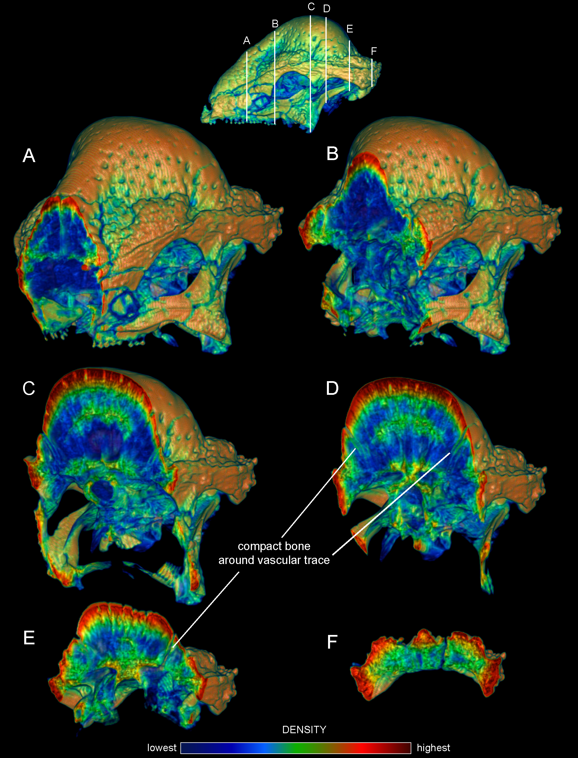
Internal densities of bone in Stegoceras validum (UA 2). Transverse CT sections from anterior (A) to posterior (F) through the cranium of Stegoceras validum (UA 2) seen in anterior oblique view. Inset CT reconstruction in lateral view (top) depicts section positions. Density and thickness of cortical bone increase towards the apex of the dome from the periphery, anteroposteriorly (B–D) and medially (C, D). Trabeculae radiate roughly perpencidular to the dome's outer surface, evident in the low-density (blue) region posterior to the orbit (B–E). Note that density of superficial bone may be inflated by beam hardening, but a dense, deep compact layer is definitively present. Dense compact bone (Hounsfield values of approximately 2000) surrounds presumed vascular traces, forming tubes that empty onto the dome surface; three of these are visible in D and E. These tubular structures recall struts within artiodactyl cranial sinuses. |
||
| Date | |||
| Source | PLoS ONE, http://www.plosone.org/article/info%3Adoi%2F10.1371%2Fjournal.pone.0021422 | ||
| Author | Eric Snively, Jessica M. Theodor | ||
| Permission (Reusing this file) |
This file is licensed under the Creative Commons Attribution 2.5 Generic license.
|
||
| Other versions | Direct link to images: http://www.plosone.org/article/slideshow.action?uri=info:doi/10.1371/journal.pone.0021422&imageURI=info:doi/10.1371/journal.pone.0021422.g001 |
File history
Click on a date/time to view the file as it appeared at that time.
| Date/Time | Thumbnail | Dimensions | User | Comment | |
|---|---|---|---|---|---|
| current | 23:56, 25 July 2016 | | 2,028 × 2,661 (3.39 MB) | FunkMonk (talk | contribs) | Cropped 2 % horizontally and 4 % vertically using CropTool with precise mode. |
| 23:55, 25 July 2016 |  | 2,063 × 2,761 (3.39 MB) | FunkMonk (talk | contribs) | {{Information |Description=Internal densities of bone in Stegoceras validum (UA 2). Transverse CT sections from anterior (A) to posterior (F) through the cranium of Stegoceras validum (UA 2) seen in anterior oblique view. Inset CT reconstruction in la... |
You cannot overwrite this file.
File usage on Commons
There are no pages that use this file.
File usage on other wikis
The following other wikis use this file:
- Usage on en.wikipedia.org
- Usage on es.wikipedia.org
- Usage on pl.wikipedia.org
- Usage on pt.wikipedia.org