File:Functional repopulation of whole-organ thymus scaffolds.webp
Jump to navigation
Jump to search
Size of this PNG preview of this WEBP file: 446 × 600 pixels. Other resolutions: 178 × 240 pixels | 357 × 480 pixels | 571 × 768 pixels | 761 × 1,024 pixels | 1,893 × 2,546 pixels.
Original file (1,893 × 2,546 pixels, file size: 1.02 MB, MIME type: image/webp)
Captions
Captions
From the study "Reconstitution of a functional human thymus by postnatal stromal progenitor cells and natural whole-organ scaffolds"
Summary[edit]
| Description |
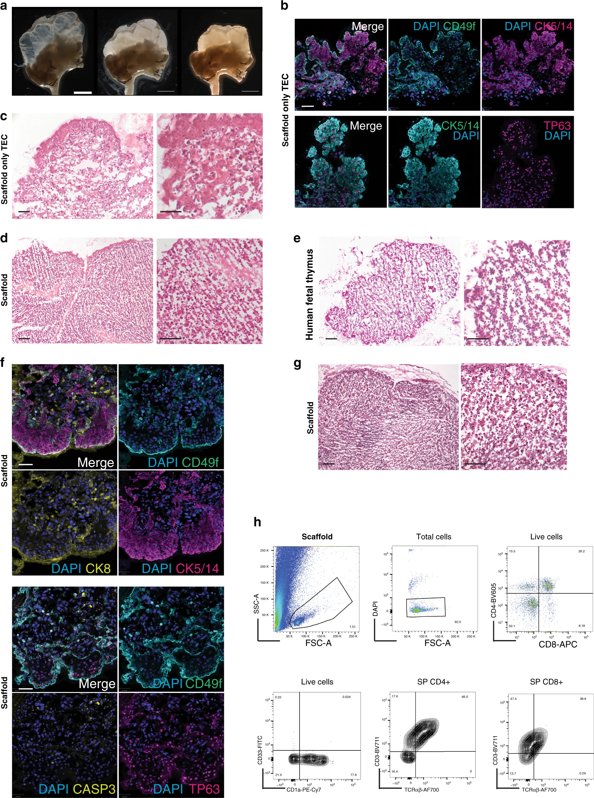
English: "a Gross microscopy representative of a thymus scaffold before (left panel), soon after injection of stromal cells (middle panel) and following 4 days of culture (right panel). Thymic lobes from empty progressively increase density and get remodelled as shown by shrinking of the scaffold and increase tissue volume (n = 60 repopulated scaffolds). Scale bar, 4 mm. b Immunofluorescence labelling of thymic epithelial cells (TEC) grown within a decellularised scaffold demonstrating presence of CK5-14+, TP63+ and CD49f+ TEC. Nuclei are stained with DAPI (n = 4 repopulated scaffolds). Scale bar, 30 μm. c H&E staining shows histology of a scaffold repopulated only with TEC and cultivated for 5 days (n = 4 repopulated scaffolds). Scale bar, 50 μm. d Haematoxyin & Eosin (H&E) staining of a scaffold repopulated with both expanded clonogenic TEC and thymic interstitial cells (TIC) and cultivated for 5 days prior to fixation and histological analysis. Stromal cells reorganise along the scaffold with a pattern similar to the one (e) observed in early (9-week post-conception, wpc) human foetal thymus (n = 4 repopulated scaffolds and n = 2 human foetal thymi). Scale bar, 100 μm. f Immunofluorescence labelling of TEC seeded together with TIC and grown within a decellularised scaffold demonstrating CK5-14+ cells localised in the subcapsular region while CK8+ cells prevalently localised in the inner regions; TP63+ TEC were mainly CD49f+. Nuclei are stained with DAPI (n = 4 repopulated scaffolds). Scale bar, 100 μm. g H&E staining of a scaffold repopulated with TEC, TIC and haematopoietic progenitors after 7 days of culture (n = 4 repopulated scaffolds). Scale bar, 100 μm. h Representative FACS analysis (n = 6 repopulated scaffold in three independent experiments) of CD45-positive population isolated from repopulated scaffold seeded with triple negative (TN, CD3−CD4−CD8−) progenitors and co-cultured for 8 days. The FSC-A, SSC-A plot displays the presence of cells as well as of debris derived from the scaffold ECM during dissociation for release of haematopoietic cells (top left panel). Viable cells were ~90% of total cells (top mid panel). TN developed within the scaffold gave rise to double positive (DP) and single positive (SP) CD4 and CD8 expressing cells (top right panel, 5000 cells). Live cells were positive for CD1a and negative for CD33 (bottom right panel). CD4 and CD8 were positive for CD3 and express TCRαβ (bottom mid and left panel)." |
| Date | |
| Source | https://www.nature.com/articles/s41467-020-20082-7 |
| Author | Authors of the study: Sara Campinoti, Asllan Gjinovci, Roberta Ragazzini, Luca Zanieri, Linda Ariza-McNaughton, Marco Catucci, Stefan Boeing, Jong-Eun Park, John C. Hutchinson, Miguel Muñoz-Ruiz, Pierluigi G. Manti, Gianluca Vozza, Carlo E. Villa, Demetra-Ellie Phylactopoulos, Constance Maurer, Giuseppe Testa, Hans J. Stauss, Sarah A. Teichmann, Neil J. Sebire, Adrian C. Hayday, Dominique Bonnet & Paola Bonfanti |
Licensing[edit]
This file is licensed under the Creative Commons Attribution 4.0 International license.
- You are free:
- to share – to copy, distribute and transmit the work
- to remix – to adapt the work
- Under the following conditions:
- attribution – You must give appropriate credit, provide a link to the license, and indicate if changes were made. You may do so in any reasonable manner, but not in any way that suggests the licensor endorses you or your use.
File history
Click on a date/time to view the file as it appeared at that time.
| Date/Time | Thumbnail | Dimensions | User | Comment | |
|---|---|---|---|---|---|
| current | 15:28, 28 January 2021 | | 1,893 × 2,546 (1.02 MB) | Prototyperspective (talk | contribs) | Uploaded a work by Authors of the study: Sara Campinoti, Asllan Gjinovci, Roberta Ragazzini, Luca Zanieri, Linda Ariza-McNaughton, Marco Catucci, Stefan Boeing, Jong-Eun Park, John C. Hutchinson, Miguel Muñoz-Ruiz, Pierluigi G. Manti, Gianluca Vozza, Carlo E. Villa, Demetra-Ellie Phylactopoulos, Constance Maurer, Giuseppe Testa, Hans J. Stauss, Sarah A. Teichmann, Neil J. Sebire, Adrian C. Hayday, Dominique Bonnet & Paola Bonfanti from https://www.nature.com/articles/s41467-020-20082-7 with U... |
You cannot overwrite this file.
File usage on Commons
There are no pages that use this file.