File:FOXJ1 expression is not sufficient to induce motile cilia formation in the chicken node.jpg
Jump to navigation
Jump to search
Size of this preview: 800 × 599 pixels. Other resolutions: 320 × 240 pixels | 640 × 479 pixels | 1,024 × 766 pixels | 1,129 × 845 pixels.
Original file (1,129 × 845 pixels, file size: 302 KB, MIME type: image/jpeg)
Captions
Captions
Add a one-line explanation of what this file represents
Summary[edit]
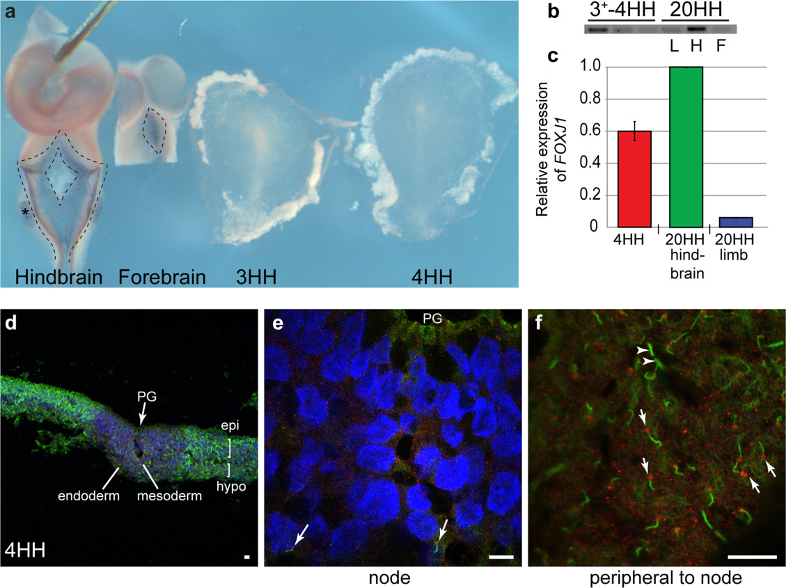
| Description | Figure 2. FOXJ1 expression is not sufficient to induce motile cilia formation in the chicken node. (a) FOXJ1 expression in the future choroid plexus of hindbrain, forebrain (outlined, arrows), and otic vesicle (asterisk) of stage 20HH embryos which precedes motile ciliogenesis, compared with stage 3HH and 4HH embryos, where no FOXJ1 can be observed (all embryos photographed underwent RNA in situ hybridization in the same tube). Red arrow indicates node, green arrow indicates primitive streak. (b) Expression of FOXJ1 by RT-PCR does show expression in stage 3HH and 4HH embryos compared to stage 20HH hindbrain (H) and forebrain (F) [negative control is stage 20HH limb bud (L)]. (c) qPCR, shows that FOXJ1 at 4HH is approximately 60% of that of the hindbrain (P = 0.000245). (d–f) Stage 4HH embryos underwent immunocytochemistry for acetylated tubulin (ciliary axoneme, green) and γ tubulin (basal body, red) to identify cilia (white arrows). Short cilia were identified in the mesoderm of the node (arrows e), while longer cilia were identified in greater abundance, in the epiblast (epi) and hypoblast (hypo) peripheral to the node (arrows f, arrowheads indicate telophase bridges which look similar to cilia but are not associated with a basal body). PG-Primitive groove; d-magnification 10×; e-magnification 50×; f-magnification 100×. |
| Date | |
| Source | https://doi.org/10.1002/dvg.22775 (2014), The chicken left right organizer has nonmotile cilia which are lost in a stage-dependent manner in the talpid3 ciliopathy. genesis, 52: 600-613. |
| Author | Stephen, L.A., Johnson, E.J., Davis, G.M., McTeir, L., Pinkham, J., Jaberi, N. and Davey, M.G. |

|
This file, which was originally posted to an external website, has not yet been reviewed by an administrator or reviewer to confirm that the above license is valid. See Category:License review needed for further instructions.
|
Licensing[edit]
This file is licensed under the Creative Commons Attribution 3.0 Unported license.
- You are free:
- to share – to copy, distribute and transmit the work
- to remix – to adapt the work
- Under the following conditions:
- attribution – You must give appropriate credit, provide a link to the license, and indicate if changes were made. You may do so in any reasonable manner, but not in any way that suggests the licensor endorses you or your use.
File history
Click on a date/time to view the file as it appeared at that time.
| Date/Time | Thumbnail | Dimensions | User | Comment | |
|---|---|---|---|---|---|
| current | 14:20, 5 May 2024 | | 1,129 × 845 (302 KB) | Rasbak (talk | contribs) | {{Information |description=Figure 2. FOXJ1 expression is not sufficient to induce motile cilia formation in the chicken node. (a) FOXJ1 expression in the future choroid plexus of hindbrain, forebrain (outlined, arrows), and otic vesicle (asterisk) of stage 20HH embryos which precedes motile ciliogenesis, compared with stage 3HH and 4HH embryos, where no FOXJ1 can be observed (all embryos photographed underwent RNA in situ hybridization in the same tube). Red arrow indicates node, green arrow... |
You cannot overwrite this file.
File usage on Commons
There are no pages that use this file.