File:Eohadrotreta zhenbaensis.jpg
Jump to navigation
Jump to search

Size of this preview: 800 × 507 pixels. Other resolutions: 320 × 203 pixels | 640 × 405 pixels | 1,024 × 649 pixels | 1,280 × 811 pixels | 2,128 × 1,348 pixels.
Original file (2,128 × 1,348 pixels, file size: 483 KB, MIME type: image/jpeg)
Captions
Captions
Add a one-line explanation of what this file represents
Summary[edit]
| Description |
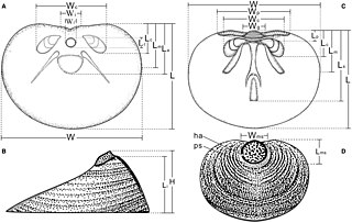
English: FIG. 2. Schematic reconstruction of Eohadrotreta zhenbaensis at ontogenetic stage T3, showing location of measurements in Figs 11–13. A–B, ventral valve; A, internal; B, lateral view. C–D, dorsal valve; C, internal; D, external view. Abbreviations: L, width; W, width; H, height of valve where not specified, and of elements: a, ventral apical process; c, cardinal muscle scars; f, pedicle foramen; g, dorsal median groove; i, ventral intertrough; m, at maximum width; ms, metamorphic shell; ps, post-metamorphic shell; p, dorsal pseudointerarea. |
| Date | |
| Source | Z. Zhang, Z. Zhang, L. E. Holmer & F. Chen: Post‐metamorphic allometry in the earliest acrotretoid brachiopods from the lower Cambrian (Series 2) of South China, and its implications. In: Palaeontology, Vol. 61, Part 2, pp. 183–207, 2018. |
| Author | Zhiliang Zhang, Zhifei Zhang, Lars E. Holmer & Feiyang Chen |
| Permission (Reusing this file) |
Open access article specified as CC BY 4.0 |
Licensing[edit]
This file is licensed under the Creative Commons Attribution 4.0 International license.
- You are free:
- to share – to copy, distribute and transmit the work
- to remix – to adapt the work
- Under the following conditions:
- attribution – You must give appropriate credit, provide a link to the license, and indicate if changes were made. You may do so in any reasonable manner, but not in any way that suggests the licensor endorses you or your use.
File history
Click on a date/time to view the file as it appeared at that time.
| Date/Time | Thumbnail | Dimensions | User | Comment | |
|---|---|---|---|---|---|
| current | 17:21, 18 October 2018 | | 2,128 × 1,348 (483 KB) | Special Circumstances (talk | contribs) | {{Information |description ={{en|1=FIG. 2. Schematic reconstruction of Eohadrotreta zhenbaensis at ontogenetic stage T3, showing location of measurements in Figs 11–13. A–B, ventral valve; A, internal; B, lateral view. C–D, dorsal valve; C, internal; D, external view. Abbreviations: L, width; W, width; H, height of valve where not specified, and of elements: a, ventral apical process; c, cardinal muscle scars; f, pedicle foramen; g, dorsal median groove; i, ventral intertrough; m, at maximu... |
You cannot overwrite this file.
File usage on Commons
There are no pages that use this file.
File usage on other wikis
The following other wikis use this file:
- Usage on de.wikipedia.org
- Usage on en.wikipedia.org
- Usage on fr.wikipedia.org
- Usage on zh.wikipedia.org