File:Deposition velocity versus cell diameters.jpg

Original file (1,355 × 1,660 pixels, file size: 206 KB, MIME type: image/jpeg)
Captions
Captions
Summary[edit]
| Description |
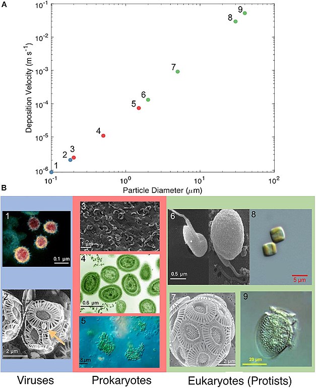
English: Deposition velocity versus cell diameters (A) Deposition velocity of varying cell diameters of viruses (blue), prokaryotes (red), and eukaryotes (green). (B) Microscopy images of microorganisms representative of the particle diameter chosen: (1) SARS-CoV-2, (2) Emiliania huxleyi virus, (3) SAR11 (Pelagibacterales), (4) Prochlorococcus, (5) Synechococcus, (6) Micromonas pusilla, (7) Emiliania huxleyi, (8) Thalassiosira sp., and (9) Dinophysis acuminata. (1–7) Approximate scale bars were added. The microscopy images in (B) were reproduced under the Creative Commons Attribution International licenses and were captured by (1) NIAID’s Rocky Mountain Laboratories in Hamilton, Montana (2) Wikimedia commons (3) Steindler et al. (2011) (4) Luke Thompson at the Sallie Chisholm Lab and Nikki Watson at Whitehead, MIT (5) Proyecto Agua at Biodiversidad virtual, Cantabria, Spain (6) Manton and Parke (1960) and courtesy of Nordic Microalgae and Aquatic Protozoa (Karlson et al., 2020) (7) Alison R. Taylor, University of North Carolina Wilmington Microscopy Facility (8) Moore et al. (2017) and (9) Plankton Net, Alfred Wegener Institute, Helmholtz Centre for Polar and Marine Research.
|
| Date | |
| Source | [1] doi:10.3389/fmicb.2021.764178 |
| Author | Alyssa N. Alsante, Daniel C. O. Thornton and Sarah D. Brooks |
| Other versions |
    |


Licensing[edit]
- You are free:
- to share – to copy, distribute and transmit the work
- to remix – to adapt the work
- Under the following conditions:
- attribution – You must give appropriate credit, provide a link to the license, and indicate if changes were made. You may do so in any reasonable manner, but not in any way that suggests the licensor endorses you or your use.
- share alike – If you remix, transform, or build upon the material, you must distribute your contributions under the same or compatible license as the original.
File history
Click on a date/time to view the file as it appeared at that time.
| Date/Time | Thumbnail | Dimensions | User | Comment | |
|---|---|---|---|---|---|
| current | 03:07, 3 December 2021 | | 1,355 × 1,660 (206 KB) | Epipelagic (talk | contribs) | Uploaded a work by Alyssa N. Alsante, Daniel C. O. Thornton and Sarah D. Brooks from [https://www.frontiersin.org/articles/10.3389/fmicb.2021.764178/full] {{doi|10.3389/fmicb.2021.764178}} with UploadWizard |
You cannot overwrite this file.
File usage on Commons
The following 8 pages use this file:
- File:Deposition velocity versus cell diameters-3.jpg
- File:Deposition velocity versus cell diameters-5.jpg
- File:Deposition velocity versus cell diameters-6.jpg
- File:Deposition velocity versus cell diameters-8.jpg
- File:Dinophysis acuminata.jpg
- File:Novel Coronavirus SARS-CoV-2.jpg
- File:Prochlorococcus marinus.jpg
- File:Virus cocco 2.jpg