File:A New Conulariid (Cnidaria, Scyphozoa) From the Terminal Ediacaran of Brazil.jpg
Jump to navigation
Jump to search
Size of this preview: 506 × 599 pixels. Other resolutions: 203 × 240 pixels | 405 × 480 pixels | 920 × 1,089 pixels.
Original file (920 × 1,089 pixels, file size: 325 KB, MIME type: image/jpeg)
Captions
Captions
A conulariid (Cnidaria) from the Ediacaran of Brazil
Summary[edit]
_From_the_Terminal_Ediacaran_of_Brazil.jpg&action=edit§ion=1)
| Description |
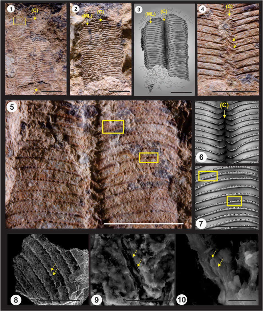
English: Paraconularia ediacara n. sp. (terminal Ediacaran, middle Tamengo Formation, upper Corumbá Group, Mato Grosso do Sul, Brazil; specimen GP-IT 2301, Geosciences Institute, University of São Paulo). 1, 2, color light photographs (both oriented with the apertural end of the fossil at the top); 1, the part, showing the two exposed partial faces and corner sulcus (indicated by the arrow labelled C) between them. The truncated apical ends of the two mostly covered faces project slightly from underneath the truncated apical ends of the two exposed faces (bottom arrow). Open yellow rectangle outlines the area from which the small fragment of periderm for SEM imaging (Figures 8–10 below) was extracted; 2, the counterpart (again with the corner indicated by an arrow labelled C, and with one of the facial midlines indicated by an arrow labelled ML); 3, schematic drawing of the part, highlighting the alternation of the nodose transverse ribs in the corner sulcus (C) and the continuation of the transverse ribs across the facial midline (ML). 4, 5, color light photographs (both oriented with the apertural end of the fossil at the top); 4, detail of the exposed corner sulcus of the counterpart. Note the pronounced adapertural deflection and alternation of the end portions of the transverse ribs within the corner sulcus (yellow arrows); 5, detail of the lower (apical) portion of the two exposed faces of the part, showing the widely spaced nodes. Upper rectangle highlights several nodes preserving the short interspace ridges in positive relief, while the lower rectangle highlights several nodes showing much shorter interspace ridges; 6, schematic drawing of a portion of the corner sulcus (C) shown in 5; 7, schematic drawing of the facial area with rectangles shown in 5; 8-10, SEM photomicrographs (secondary electron mode) of a small fragment of the periderm; 8, exterior surface of the periderm, showing several transverse ribs, widely spaced nodes, and very short, spine-like interspace ridges (pointing toward the apertural end of the periderm, yellow arrows); 9, detail of the fragment shown in 8, with canyon-like fractures exposing the edges of several microlamellae (yellow arrows); 10, detail of one of the fractures shown in 9 and exposing microlamellae (yellow arrows). Scale bar: 5 to 8 mm (Figures 1–3, 5); 7 mm (Figure 4); 5 mm (Figures 6, 7); 3 mm (Figure 8); 40 μm (Figure 9); 5 μm (Figure 10). |
| Date | |
| Source | https://www.frontiersin.org/articles/10.3389/feart.2022.777746/full |
| Author | Juliana M. Leme, Heyo Van Iten, and Marcello G. Simões |
Licensing[edit]
_From_the_Terminal_Ediacaran_of_Brazil.jpg&action=edit§ion=2)
This file is licensed under the Creative Commons Attribution-Share Alike 4.0 International license.
- You are free:
- to share – to copy, distribute and transmit the work
- to remix – to adapt the work
- Under the following conditions:
- attribution – You must give appropriate credit, provide a link to the license, and indicate if changes were made. You may do so in any reasonable manner, but not in any way that suggests the licensor endorses you or your use.
- share alike – If you remix, transform, or build upon the material, you must distribute your contributions under the same or compatible license as the original.
File history
Click on a date/time to view the file as it appeared at that time.
| Date/Time | Thumbnail | Dimensions | User | Comment | |
|---|---|---|---|---|---|
| current | 03:05, 23 December 2022 | | 920 × 1,089 (325 KB) | Fossiladder13 (talk | contribs) | Uploaded a work by Juliana M. Leme, Heyo Van Iten, and Marcello G. Simões from https://www.frontiersin.org/articles/10.3389/feart.2022.777746/full with UploadWizard |
You cannot overwrite this file.
File usage on Commons
There are no pages that use this file.
File usage on other wikis
The following other wikis use this file:
- Usage on en.wikipedia.org
_From_the_Terminal_Ediacaran_of_Brazil.jpg&oldid=776446331)