File:41598 2015 Article BFsrep14735 Fig1a-Glaucocystis geitleri.jpg
Jump to navigation
Jump to search

Size of this preview: 713 × 600 pixels. Other resolutions: 285 × 240 pixels | 571 × 480 pixels | 776 × 653 pixels.
Original file (776 × 653 pixels, file size: 95 KB, MIME type: image/jpeg)
Captions
Captions
Add a one-line explanation of what this file represents
Summary[edit]
| Description |
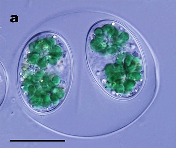
English: Differential interference contrast microscopy of a vegetative cell of the coccoid glaucophyte species “Glaucocystis geitleri” strain SAG 229-1. Scale bar, 20 μm. Note that immobile vegetative cells are enclosed by a cell wall within an expanded mother cell wall. |
| Date | |
| Source |
Fig. 1a at https://www.nature.com/articles/srep14735 Ultra-high voltage electron microscopy of primitive algae illuminates 3D ultrastructures of the first photosynthetic eukaryote. In: Scientific Reports volume 5, Article number: 14735. doi:10.1038/srep14735 |
| Author | Toshiyuki Takahashi, Tomoki Nishida, Chieko Saito, Hidehiro Yasuda, Hisayoshi Nozaki |
| Other versions |
Licensing[edit]
This file is licensed under the Creative Commons Attribution-Share Alike 4.0 International license.
- You are free:
- to share – to copy, distribute and transmit the work
- to remix – to adapt the work
- Under the following conditions:
- attribution – You must give appropriate credit, provide a link to the license, and indicate if changes were made. You may do so in any reasonable manner, but not in any way that suggests the licensor endorses you or your use.
- share alike – If you remix, transform, or build upon the material, you must distribute your contributions under the same or compatible license as the original.
File history
Click on a date/time to view the file as it appeared at that time.
| Date/Time | Thumbnail | Dimensions | User | Comment | |
|---|---|---|---|---|---|
| current | 07:52, 11 October 2021 | | 776 × 653 (95 KB) | Ernsts (talk | contribs) | Uploaded a work by Toshiyuki Takahashi, Tomoki Nishida, Chieko Saito, Hidehiro Yasuda, Hisayoshi Nozaki from Fig. 1-3 at https://www.nature.com/articles/srep14735 Ultra-high voltage electron microscopy of primitive algae illuminates 3D ultrastructures of the first photosynthetic eukaryote Scientific Reports volume 5, Article number: 14735. doi:doi.org/10.1038/srep14735 50px|class=noviewer with UploadWizard |
You cannot overwrite this file.
File usage on Commons
The following page uses this file: